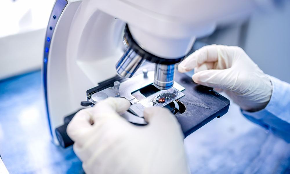
Association’s New Database Puts Anatomy Under a Virtual Microscope
The American Association of Anatomists is pooling together the virtual microscopic samples of research facilities around the country with its new Virtual Microscopy Database. The database idea was formulated by an interest group within AAA.
In the past, it was hard to get a close look at the intricacies of human anatomy without a microscope. Even as microscopes gave way to computers, it was hard to find high-quality digital slides without a lot of work.
But a $50,000 initiative from the American Association of Anatomists could change that. Last week, AAA launched its first Virtual Microscopy Database (VMD), making it possible for researchers to get an up-close look at a wide variety of virtual tissue slides with the click of a button.
The concept came about from AAA’s Digital Histology Interest Group, noted the University of Colorado’s Lisa Lee, Ph.D. The group, she says, highlighted the shared problems that anatomy and histology scholars often dealt with.
“In our discussions, many of us lamented the fact that our digital slide collection was limited in diversity, so we were each on the hunt for different slides, rare tissues, or something each of us lacked,” Lee explained. “VMD meets these needs all in one place and helps us build a community for not only resource sharing, but also networking and collaboration.”
The database isn’t intended for the student population but is instead designed for researchers and institutions to share their microscopic samples. Researchers approved for the program will be expected to share their anatomical samples with others taking part.
The approach helps improve resourcing across the board as research facilities increasingly move away from traditional microscope samples in favor of virtual microscopy, which is used in many schools. The University of Michigan’s Michael Hortsch, Ph.D., notes that the approach helps schools keep down costs from maintaining older equipment while raising the overall quality.
“More and more schools forgo the expensive purchase and maintenance of microscopes and glass histology slides that can break and instead use virtual microscopy with computers to teach students the microscopic structure and function of cells, tissues, and organs,” Hortsch noted. “As no single slide collection is complete and flawless, pooling image collections from many different schools and making them available to all histology educators and researchers in the form of the VMD electronic database is a perfect solution.”
(iStock/Thinkstock)


Comments